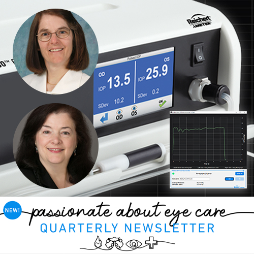
By Carol Toris, PhD and Cynthia J. Roberts, PhD
The Model 30™ Pneumatonometer is in a class all its own. Developed by Durham and Langham in the mid-1960s, the Pneumatonometer, like Goldmann Applanation Tonometry, is a fixed area/variable force applanation tonometer. However, whereas the Goldmann measurement is “static” in nature, the Pneumatonometer utilizes a dynamic methodology, which makes this device unique.
How Does It Work?
Applanation is achieved via compressed air that flows through flexible tubing into a handheld probe. The probe features a piston shaft, actuated by the air flow, with a silicone-membrane covered contact-tip. The flowing air escapes through perforations in the back of the tip. When the tip is applied to the cornea, back-pressure builds in the system, which is recorded by a pressure transducer. The piston shaft is floating on an “air bearing”, which enables recording of the systolic and diastolic ocular pressure. The average of these pulse pressures is reported as the IOP. IOP measurements can be made with the subject in seated or supine positions which is useful if measurements are needed at night in addition to day.1,2 The subject need not sit in front of a slit lamp as required by Goldmann Tonometry. Measurements also can be made in research animals including cats,3 dogs,4 rabbits,5 and monkeys.6
Measurements of Ocular Pulse Amplitude
Intraocular pressure (IOP) is not a static quantity, as is often assumed, but rather varies with each heartbeat. As the vessels in the choroid fill in a pulsatile pattern that directly reflects cardiac output, the IOP increases and then decreases, thus producing a pulsatile ocular pressure signal. The peak to valley amplitude is termed the ocular pulse amplitude (OPA) and the Pneumatonometer methodology facilitates its quantification. OPA is a function of both ocular blood volume and ocular rigidity.7 The greater the ocular rigidity, the greater the ocular pulse and OPA increases as IOP increases in a linear relationship. In addition, the greater the pulsatile ocular blood volume (POBV), the greater the OPA. However, as IOP increases, POBV decreases, so there is an opposite relationship between OPA and IOP which is positive, and POBV vs IOP which is negative. Our research finds that higher IOP with lower OPA indicates lower ocular blood volume.8 POBV is lower in the eye with worse disease in asymmetric open angle glaucoma.9 Calculation of POBV requires a measurement of axial length in order to estimate ocular rigidity. However, a comparison between POBV and OPA/IOP (diastolic) showed a strong linear relationship which means POBV can be estimated with knowledge of OPA and IOP, both of which can easily be measured with the Model 30 Pneumatonometer.
Measurements of Outflow Facility
The primary reason for the intraocular pressure elevation in most forms of glaucoma is a reduction in the facility of outflow through the trabecular meshwork. For those who study or treat glaucoma, outflow facility (C) is a crucial parameter of aqueous humor dynamics to investigate. In humans, it can be done by a procedure called tonography. Adding a 10-gram weight to the probe shaft of the Pneumatonometer hand piece provides tonography measurements. While the patient is in a supine position, the weighted probe is applied to the eye for two to four minutes and IOP is measured continuously. A series of complex equations are applied to calculate C. The Model-30 Pneumatonometer tonography module has been used in numerous clinical studies to assess IOP and C. These studies have included effects of drugs, surgical procedures, aging, 24 hour fluctuations, and differences related to ethnic background. The Pneumatonometer has contributed to the improved understanding of glaucoma and its treatments.
Digitizing the Data
For decades, the Pneumatonometer featured a strip-chart printer that recorded the measurement data graphically, which is neither friendly for the modern digital era, nor efficient. Costly replacement paper rolls and the risk of losing these small strips of paper were a pain point for many doctors and clinicians since the data could not be digitized or exported for analysis. To bring this unique device into the modern age, a much-awaited upgrade was recently released.
Responding to requests from users, Reichert introduced an updated version of the Model 30 Pneumatonometer that features USB export to a new PC application with real-time digital monitoring and recording of the signal and measurement data. In addition to viewing the signal and data in the PC application, all >4000 IOP data points can be exported to an analysis program of the investigator’s choosing, such as Microsoft Excel, enabling detailed analysis of data and graphing of results. One must have a good knowledge of tonography and its formulas to calculate outflow facility, but the new functionality is a big step towards this goal. This welcomed update expands the utility of the Model 30 Pneumatonometer and ensures that it will remain relevant and useful to ophthalmic researchers and clinicians now and into the future.
References
1. Liu H, Fan S, Gulati V, et al. Aqueous humor dynamics during the day and night in healthy mature volunteers. Arch Ophthalmol. Mar 2011;129(3):269-75. doi:10.1001/archophthalmol.2011.4
2. Fan S, Hejkal JJ, Gulati V, Galata S, Camras CB, Toris CB. Aqueous humor dynamics during the day and night in volunteers with ocular hypertension. Arch Ophthalmol. Sep 2011;129(9):1162-6. doi:10.1001/archophthalmol.2011.226
3. Toris CB, Yablonski ME, Wang YL, Hayashi M. Prostaglandin A2 increases uveoscleral outflow and trabecular outflow facility in the cat. Experimental eye research. Dec 1995;61(6):649-57. doi:10.1016/s0014-4835(05)80015-6
4. Toris CB, Lane JT, Akagi Y, Blessing KA, Kador PF. Aqueous flow in galactose-fed dogs. Experimental eye research. 2006/10/01/ 2006;83(4):865-870. doi:https://doi.org/10.1016/j.exer.2006.04.005
5. Zhan GL, Toris CB, Camras CB, Wang YL, Yablonski ME. Bunazosin reduces intraocular pressure in rabbits by increasing uveoscleral outflow. J Ocul Pharmacol Ther. Jun 1998;14(3):217-28. doi:10.1089/jop.1998.14.217
6. Toris CB, Zhan GL, Wang YL, et al. Aqueous humor dynamics in monkeys with laser-induced glaucoma. J Ocul Pharmacol Ther. Feb 2000;16(1):19-27. doi:10.1089/jop.2000.16.19
7. Perkins ES. The ocular pulse. Curr Eye Res. 1981;1(1):19-23. doi:10.3109/02713688109019968
8. Somogye R, Roberts CJ, Spoerl E, Pillunat KR, Pillunat LE, Small R. Predicting Pulsatile Ocular Blood Volume Non-invasively. Invest Ophthalmol Vis Sci. 2021;62(8):547-547.
9. Harvey D, Roberts CJ, Mahmoud A, Fleming G. Comparisons of Biomechanical Metrics between eyes of patients with Asymmetric Glaucoma and Symmetric Glaucoma. Invest Ophthalmol Vis Sci. 2022;63(7):2384 – A0187-2384 – A0187.